Bone Cross Section Diagram - Compact Bone Diagram Cell Diagram Skeletal System Anatomy Bones Basic Anatomy And Physiology - Diagram with articular cartilage, marrow, spongy bone, medullary cavity, endosteum, diaphysis, and periosteum.. They build the entire picture, improve your understanding, consolidate the information and facilitate recall. Vector illustration scheme of bone cross section. Bone cross section diagram ipad folio cases. Hi all, i have uploaded a new medical animation tutorial. Internal structure of the dicotyledonous stem by openstax.
For example, to read this diagram literally, since the cartilage can be seen inside the cutaway section of bone, it. They build the entire picture, improve your understanding, consolidate the information and facilitate recall. Vector illustration scheme of bone cross section. Cross section of bone diagram. This is a short tutorial using blender 2.8 that shows how to create a bone cross section and using images to create the textures.
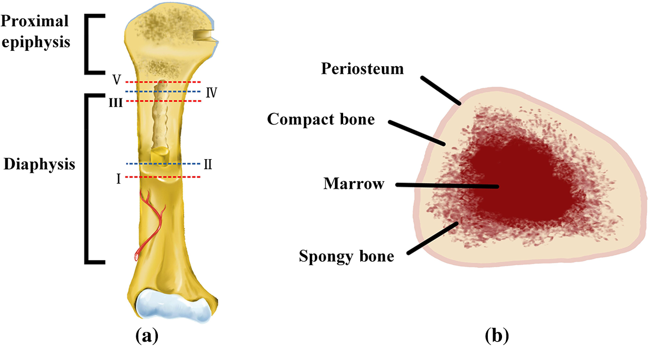
Application Of Continuous Wave Terahertz Computed Tomography For The Analysis Of Chicken Bone Structure from www.spiedigitallibrary.org Comprar este vector de stock y explorar vectores similares en adobe stock. Vector illustration scheme of bone cross section. Diagram with articular cartilage, marrow, medullary cavity and periosteum. Cross section through middle metacarpal bones of vector. Bone is found in the shafts of long bone and consists of various cylindrical units named as haversian system 47. Explaned distal and proximal epiphysis. The centroidal locations of common cross sections are well documented, so it is typically not necessary to calculate the location with the equations above. (b) in this micrograph of the osteon, you can clearly see the concentric lamellae and central canals.
Download 130+ royalty free bone cross section vector images.
Bone cross section diagram ipad folio cases. There are trabeculae in spongy bone which gives its sponge like appearance. Cross section of bone diagram. The centroidal locations of common cross sections are well documented, so it is typically not necessary to calculate the location with the equations above. Skin anatomy diagram description illustration skin stock.
File Bone Cross Section Svg Wikimedia Commons Bone Marrow Yellow Bone Marrow Cancellous Bone from i.pinimg.com Comprar este vector de stock y explorar vectores similares en adobe stock. Related posts of bone cross section labeled. Healthy tooth diagram isolated on white background vector. (b) in this micrograph of the osteon, you can clearly see the concentric lamellae and central canals. As shown in figure 2. Hi all, i have uploaded a new medical animation tutorial. Vector illustration scheme of bone cross section. Tooth anatomy, medical labeled cross section chart with enamel, dentin, pulp, gingiva, blood vessels and nerves.
Jump to navigation jump to search.
Compact bone is the outer layer and the spongy bone forms the inner layer. As shown in figure 2. Related posts of cross section of human body organs skeleton bones diagram. Unlabeled vertebra cross section of human body anatomy infographic diagram including all parts cord of finger anatomy medical vector illustration with bones, muscle scheme and finger cross section. Spinal cord spinal column anatomy information myvmc. Bone cross section diagram ipad folio cases. There are trabeculae in spongy bone which gives its sponge like appearance. Explaned distal and proximal epiphysis. Each system contains haversian canals surrounded by concentric lamellae of bone tissue 48. In a cross section of a bone we can see two types of bone tissue: Vector illustration scheme of bone cross section. (micrograph provided by the regents of university of michigan. Hope you enjoy and please.
Related posts of cross section of human body organs skeleton bones diagram. The centroidal locations of common cross sections are well documented, so it is typically not necessary to calculate the location with the equations above. Related posts of bone cross section labeled. Tooth anatomy, medical labeled cross section chart with enamel, dentin, pulp, gingiva, blood vessels and nerves. Vector illustration scheme of bone cross section.
A P 1 Lab Long Bone Cross Section Flashcards Quizlet from o.quizlet.com Related posts of bone cross section labeled. See labeled cross sections of the human body now at spinal cord crosssection images stock photos vectors shutterstock. Diagram with articular cartilage, marrow, spongy bone, medullary cavity, endosteum, diaphysis, and periosteum.: Vector illustration scheme of bone cross section. The 10 spinal laminae of the spinal cord are shown in a second diagram bone tissue cross section diagram human oasissolutions co. For example, to read this diagram literally, since the cartilage can be seen inside the cutaway section of bone, it. Human anatomy for muscle, reproductive, and skeleton. The centroidal locations of common cross sections are well documented, so it is typically not necessary to calculate the location with the equations above.
(micrograph provided by the regents of university of michigan.
(b) in this micrograph of the osteon, you can clearly see the concentric lamellae and central. Cross section of bone diagram. Internal structure of the dicotyledonous stem by openstax. As shown in figure 2. Two prominent grooves or sulci run along its length. Jump to navigation jump to search. They build the entire picture, improve your understanding, consolidate the information and facilitate recall. (b) in this micrograph of the osteon, you can clearly see the concentric lamellae and central canals. Tooth anatomy, medical labeled cross section chart with enamel, dentin, pulp, gingiva, blood vessels and nerves. From wikimedia commons, the free media repository. Spinal cord spinal column anatomy information myvmc. Jump to navigation jump to search. Vector illustration scheme of bone cross section.
Healthy tooth diagram isolated on white background vector bone cross section. Tooth anatomy, medical labeled cross section chart with enamel, dentin, pulp, gingiva, blood vessels and nerves.


0 Komentar